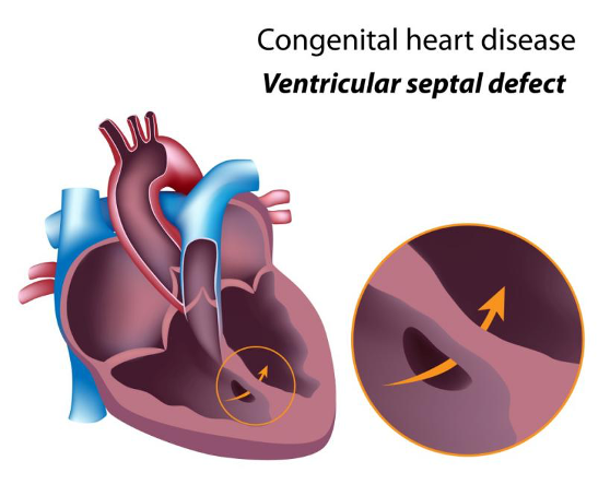
VSD is a form of hole in the wall of heart that is situated between two lower chambers of the heart known as ventricular septum. This is a congenital heart disease (CHD), meaning by present since birth.
Due to this hole, blood from left sided lower chamber (Left ventricle, pure blood mixes with impure blood (right sided lower chamber, right ventricle), resulting into overflow of blood of both lungs resulting into heart failure of the child.
Ventricular septal defect (VSD) is one of the commonest child heart diseases that are seen at birth, but adults too may suffer from it following an acute heart attack.
What is the cause of Ventricular Septal Defect (VSD causes)?
Congenital ventricular septal defect (congenital VSD) occurs during development of fetus heart in first 12 weeks of pregnancy. If development of heart goes wrong in first 12 weeks of pregnancy, it can results in a hole in the ventricular septum.
Sometimes, VSD may be caused due to genetic and environmental factors. For instance, if you have a family history of genetic conditions like Down syndrome or congenital heart diseases, then your child may be at risk of developing a Ventricular Septal Defect.
Types of Ventricular Septal Defect (VSD):
- Peri-membranous VSD: This is most common type of VSD, situated in upper part of ventricular septum.
- Inlet VSD
- Outlet VSD
- Muscular VSD: This type of VSD is generally closed by itself automatically.

Dr Gaurav Agrawal is a Pediatric Cardiologist (Child and Fetal heart Specialist). Dr Agrawal is a pediatrician who has specialized training in the treatment of various child heart related problems (neonates to 18 years of age). He is also having a vast experience in fetal echocardiography.
He is one of the very few pediatricians in India who practices pediatric cardiology exclusively. Cardiac problems in children are common in children, sometimes life threating and often not diagnosed properly. Early diagnosis and timely treatment is very important. Areas of specific interest include child echocardiography including fetal echocardiography, diagnostic and therapeutic cardiac catheterization (pediatric cardiac holes closure by device, opening of obstructed cardiac valves by balloons etc).


